Location: Home >> Detail
J Psychiatry Brain Sci. 2026;11(5):e260017. https://doi.org/10.20900/jpbs.20260017
 ,
Chongben Zhang 2 ,
Wenxin Bian 3 ,
Lanye Wang 4 ,
Ya-Ke Wu 3,5,*
1
2
3
4
5
*
Background: Binge eating and loss-of-control (LOC) eating behaviors may persist or recur after bariatric surgery, but the underlying biological mechanisms remain unclear. This pilot study aimed to (1) examine changes in binge eating, LOC eating, appetite hormones, and appetite-related gene expression before (T1) and 3 months after bariatric surgery (T2) and (2) test whether these biological markers were associated with post-surgical binge or LOC eating. Methods: Binge eating and LOC eating were assessed using the Binge Eating Scale and the Loss of Control Over Eating Scale. Fasting blood samples were analyzed for leptin, ghrelin, and glucagon-like peptide-1 (GLP-1) levels, as well as the expression levels of the LEP, LEPR, GHRL, GLP1R, and GCG genes. Pre- to post-surgery changes were examined using Wilcoxon signed-rank tests. Generalized linear regression was used to test associations with post-surgical disordered eating. Results: Participants (total n = 21) showed significant decreases in BMI, binge eating, and LOC eating from T1 to T2. Leptin and GLP-1 levels significantly decreased at T2. No biomarkers were significantly associated with disordered eating. Conclusions: The findings highlight significant reductions in BMI, disordered eating behaviors, and leptin and GLP-1 levels following bariatric surgery. These preliminary results warrant further investigation into the biological mechanisms underlying post-surgical disordered eating in larger, more diverse cohorts with longer follow-up.
BES, Binge Eating Scale; GLP-1 , glucagon-like peptide-1; IQR, interquartile range; LOC, loss of control; mRNA, messenger RNA; RYGB, Roux-en-Y gastric bypass
Binge eating and loss of control (LOC) eating frequently occur or persist following bariatric surgery, with post-surgical prevalence estimates ranging from 3% to 37% for binge eating and 8% to 50% for LOC eating [1]. Longitudinal studies have shown that although the severity of these behaviors might decrease initially after surgery, their severity often increases significantly after the first postoperative year [2–4]. These disordered eating behaviors are clinically significant, as they are associated with poorer weight loss outcomes and an elevated risk of weight regain [1]. Despite their clinical relevance, the biological mechanisms contributing to disordered eating following bariatric surgery remain poorly understood.
Appetite-related hormones, such as leptin (a satiety-promoting hormone primarily produced by adipocytes), ghrelin (a hunger-stimulating hormone primarily produced by the stomach), and glucagon-like peptide-1 (GLP-1; a satiety-promoting hormone primarily produced by intestinal L cells), are key regulators of food intake and have been implicated in the pathophysiology of eating disorders [5–7]. Hormonal changes following bariatric surgery have been documented, such as decreased ghrelin levels, increased GLP-1 levels, and mixed findings for leptin levels [8–10], yet the association between these hormone profiles and post-surgical disordered eating behaviors remains poorly understood. In addition to the circulating levels of appetite-related hormones, the expression of their corresponding genes, including leptin (LEP), leptin receptor (LEPR), ghrelin (GHRL), glucagon-like peptide-1 receptor (GLP1R), and glucagon (GCG), may also contribute to the development or persistence of disordered eating behaviors following bariatric surgery. Changes in gene expression are typically assessed by measuring changes in messenger RNA (mRNA) levels, which may result in altered protein production (upregulation = increased mRNA/protein; downregulation = decreased mRNA/protein) [11]. For example, although circulating ghrelin levels are initially reduced after Roux-en-Y gastric bypass (RYGB) because the surgery bypasses the distal stomach (where most ghrelin-producing cells are located) [8], increased expression of ghrelin-related genes has been observed within 3 months post-surgery [12], suggesting a potential compensatory increase in ghrelin production capacity. Upregulated ghrelin-related gene expression after surgery may increase ghrelin release and enhance signaling to brain regions that regulate appetite and reward, thereby heightening vulnerability to binge or LOC eating post-surgery [13].
While these findings support a plausible biological mechanism, it remains unknown whether post-surgical changes in appetite hormone levels and their gene expression directly contribute to the development or recurrence of binge eating and LOC eating. To address this gap, this pilot study aimed to 1) evaluate changes in binge eating, LOC eating, appetite hormone levels (i.e., leptin, ghrelin, and GLP-1), and gene expression of appetite-related genes (i.e., LEP, LEPR, GHRL, GLP1R, and GCG) before and 3 months after bariatric surgery; and 2) examine whether pre- or post-surgical biological markers, as well as the change between two time points, are associated with binge or LOC eating following surgery. The rationale for the 3-month post-surgery follow-up was based on prior evidence indicating increased expression of appetite-related genes during the early postoperative period [12]. Findings from this study will provide preliminary evidence on the potential biological mechanisms of disordered eating after bariatric surgery, which will inform future larger-scale research exploring the metabolic pathways that influence disordered eating behaviors following bariatric procedures.
Participants were recruited using social media and the University of North Carolina at Chapel Hill (UNC-Chapel Hill) Research for Me website. Flyers were provided to participants with an interest in sharing the study information. Eligible individuals were at least 18 years old, resided in North Carolina, could read and write English at a sixth-grade level or higher, were candidates for any bariatric surgery scheduled within 12 months, had no current or past use of weight loss medications, and had no history of coagulation disorders due to the blood sample collection required for this study. Eligibility screening was conducted online via REDCap and followed by informed consent and self-report questionnaires. Eligible participants completed two in-person visits at home or in the laboratory at baseline (T1, pre-surgery) and 3-month follow-up (T2, post-surgery), during which a trained research assistant and the principal investigator collected anthropometric data and obtained fasting blood samples. At 3 months, participants were emailed a post-surgery online survey to complete and were scheduled for a second in-person visit. Progressive incentives and free parking were provided to encourage retention. This study was approved by the Institutional Review Board (IRB) at UNC-Chapel Hill (IRB number: 22-3176). In total, 21 participants completed the pre-surgery survey, and 11 also underwent in-person anthropometric measurements and blood sampling. At the 3-month follow-up, 10 participants completed the post-surgery survey, and 9 provided anthropometric measurements and blood samples.
Measurements Demographic VariablesThe participants self-reported their age, sex, race, and ethnicity when completing online surveys.
Clinical OutcomesAt both time points, weight and height were each measured twice and averaged. BMI was then calculated as weight in kilograms divided by height in meters squared (kg/m2). Binge eating at T1 and T2 was assessed with the Binge Eating Scale (BES), a 16-item self-report questionnaire [14]. Each item was rated on a Likert scale ranging from 0 to 3 or from 0 to 4, with the total score ranging from 0 to 46. Higher scores reflect more severe binge eating. Scores below 17 indicate non-binge eating, scores from 18 to 26 indicate moderate binge eating, and scores above 27 indicate severe binge eating; these cutoffs have been validated in a bariatric surgery-seeking population [15]. In the current study, the BES had a Cronbach’s alpha of 0.96 at T1 and 0.83 at T2. LOC eating at T1 and T2 was assessed using the Loss of Control Over Eating Scale (LCOES) [16], a 24-item self-reported questionnaire. Each item was rated on a Likert scale ranging from 1 to 5, with the total score ranging from 24 to 120. Higher scores reflect more severe LOC eating. In the current study, the Cronbach’s alpha of the LCOES was 0.86 at both time points.
Hormone MeasuresFasting whole blood samples (5 mL) were collected at T1 and T2 to quantify circulating levels of appetite-related hormones, including leptin, ghrelin, and GLP-1. Hormone concentrations were analyzed at the UNC-Chapel Hill Biobehavioral Laboratory using the Luminex FLEXMAP 3D multiplexing platform (Thermo Fisher Scientific; Waltham, MA, USA) and a human metabolic hormone magnetic bead panel kit (MilliporeSigma; Burlington, MA, USA) following the manufacturer’s instructions. Briefly, samples, standards, and controls were incubated overnight at 4 °C with primary antibody-conjugated beads. After washing, beads were incubated with detection antibodies for 1 hour at room temperature, followed by incubation with the fluorescent conjugated streptavidin-phycoerythrin for 30 min at room temperature. After a final wash, fluorescent signals were detected using the Luminex platform. Hormone concentrations were calculated from standard curves generated by the instrument’s built-in software.
Gene ExpressionFasting whole blood samples (5 mL) were collected at T1 and T2 to assess gene expression. Gene expression assays were conducted at the UNC-Chapel Hill Biobehavioral Laboratory using QuantStudio 12K Flex. Total RNA was extracted from blood using a Tempus Spin RNA Isolation Reagent Kit (Applied Biosystems; Waltham, MA, USA). RNA concentration and purity were assessed with a NanoDrop spectrophotometer. cDNA was synthesized using a high-capacity cDNA reverse transcription kit (Applied Biosystems), using 45 ng/sample total RNA. The cDNA concentration was determined with a NanoDrop spectrophotometer. TaqMan probes and master mix (Applied Biosystems) were used for RT-PCR with a total reaction volume of 10 µL (5 µL master mix, 2 µL cDNA, 0.5 µL probe, 2.5 µL H2O). Triplicates were performed, and the mean cycle threshold (Ct) value across technical replicates was used for analysis. For each sample, target gene expression was normalized to the expression of the internal control, actin, by calculating ΔCt as the target gene Ct minus the actin Ct. Lower ΔCt values indicate higher relative target gene expression, whereas higher ΔCt values indicate lower relative expression. Changes in relative gene expression from pre- to post-surgery were assessed using ΔΔCt, calculated as post-surgery ΔCt minus pre-surgery ΔCt.
Statistical AnalysisDescriptive statistics were calculated for demographic, clinical, hormonal, and gene expression variables, with medians and interquartile ranges (IQRs) reported for continuous variables and frequencies and percentages reported for categorical variables. Changes from pre- to post-surgery in clinical outcomes (BMI, binge eating, and LOC eating), hormone levels (leptin, ghrelin, and GLP-1), and gene expression (ΔCt for LEP, LEPR, GHRL, GLP1R, and GCG) were examined using the Wilcoxon signed-rank test.
Generalized linear regression models were used to examine the associations of appetite-related hormone concentrations and gene expression levels with post-surgical binge eating and LOC eating. Separate models were conducted for each biomarker at three time points: pre-surgery, post-surgery, and change from pre- to post-surgery. For regression analyses of gene expression at the pre- and post-surgery time points, −ΔCt was used, with higher values indicating higher relative gene expression. Change in gene expression was represented as −ΔΔCt, with positive values indicating increased expression and negative values indicating decreased expression from pre- to post-surgery. Models for post-surgical binge eating were adjusted for pre-surgical BMI and pre-surgical binge eating, while models for post-surgical LOC eating were adjusted for pre-surgical BMI and pre-surgical LOC eating. No corrections were applied for multiple comparisons due to the exploratory nature of this pilot study. Because most ghrelin values were below the lower limit of detection, only a small number of complete observations were available for pre-surgery, post-surgery, and change analyses, respectively (n = 5, 4, and 1); therefore, paired tests and adjusted regression models could not be conducted for ghrelin concentrations.
Table 1 presents the demographic, clinical, and laboratory characteristics of the study sample. Participants had a median age of 46 years (IQR: 41–53), and the majority were female, Caucasian, and non-Hispanic. Following bariatric surgery, participants showed a significant reduction in BMI (p < 0.01), as well as decreased median scores for binge eating (p = 0.03) and LOC eating (p = 0.02) compared to pre-surgery levels. Among the appetite hormones, leptin (p = 0.04) and GLP-1 (p = 0.01) levels significantly decreased after surgery. No statistically significant change was observed in the expression levels of appetite-related genes.
Table 2 presents the results of the generalized linear regression analyses. No significant associations were observed between hormone or gene expression levels and disordered eating outcomes following surgery. Figure 1 summarizes the study design and changes in key behavioral and physiological outcomes.
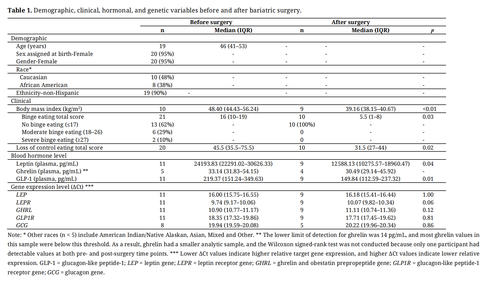 Table 1.
Demographic, clinical, hormonal, and genetic variables before and after bariatric surgery.
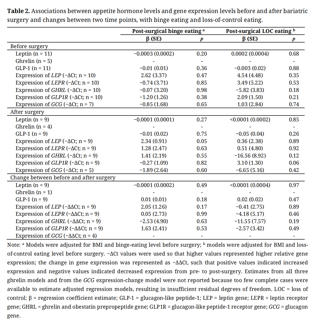 Table 2.
Associations between appetite hormone levels and gene expression levels before and after bariatric surgery and changes between two time points, with binge eating and loss-of-control eating.
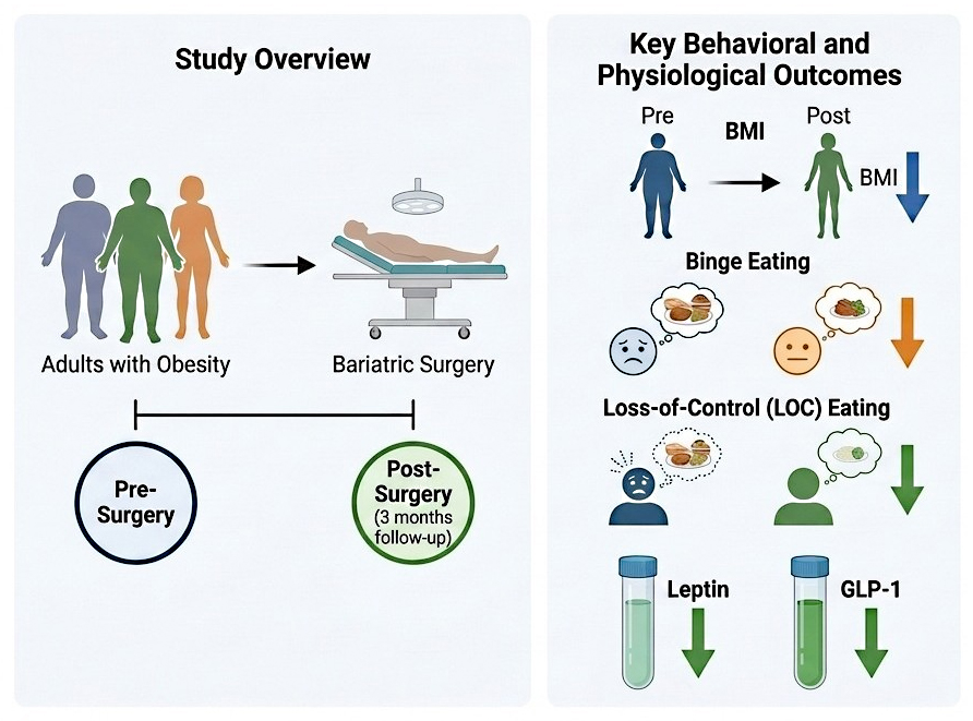 Figure 1. Study design and key findings.
The current pilot study examined pre- to post-surgical changes in disordered eating behaviors, appetite-related hormones, and gene expression in a sample of adults undergoing bariatric surgery. Participants exhibited significant reductions in BMI, binge eating, and LOC eating following surgery, which is consistent with previous research demonstrating short-term improvements in disordered eating behaviors post-surgery [2]. Leptin and GLP-1 levels significantly decreased from baseline to follow-up. The decrease in leptin is consistent with previous studies [8,9]. For GLP-1, prior studies following bariatric surgery have shown different patterns depending on the timing of measurement, with postprandial GLP-1 responses consistently increasing after surgery, whereas fasting GLP-1 levels are more variable [8–10]. In contrast, no significant pre- to post-surgical changes were observed in the expression of the appetite-related genes examined. However, given the small number of participants with available gene expression data, these findings should be considered preliminary and interpreted with caution.
Although GLP-1 signaling is known to promote satiety and reduce the hedonic drive for food, and pharmacologic GLP-1 receptor agonists have shown promise in reducing binge eating [17], we did not observe a statistically significant association between GLP1R expression and post-surgical disordered eating. Given the small sample size and exploratory nature of this pilot study, these findings should be interpreted cautiously. Furthermore, GLP1R expression and LOC eating were assessed at the same postoperative time point in our study. Future longitudinal studies with larger samples and extended follow-up are needed to determine whether GLP1R expression is associated with LOC eating post-surgery.
Our findings are consistent with previous research showing improvements in disordered eating behaviors and changes in appetite-related hormone levels following bariatric surgery. Several limitations should be noted. First, the sample size was small, which may have limited power to detect associations between biological markers and disordered eating behaviors. The follow-up duration was relatively short. Although persistent changes in appetite-related hormone levels after weight loss have been documented over longer follow-up periods [18–20], the long-term trajectories of gene expression after bariatric surgery remain unclear. Furthermore, leptin, ghrelin, and GLP-1 are primarily adiposity- and gut-derived metabolic hormones released from tissue-specific cells. Ideally, assessment of their gene expression would be performed in the relevant tissues, such as subcutaneous adipose tissue or gastric cells, to best reflect local transcriptional activity. However, owing to funding constraints, we were only able to measure gene expression from whole blood samples, which may not fully capture tissue-specific regulation and could limit the interpretability of our findings regarding the relationship between hormone gene expression and eating behaviors. In particular, given the potential relevance of GLP-1 signaling to eating behaviors, future studies in animal models could compare GLP1R mRNA expression in peripheral blood with expression in GLP-1-responsive tissues, including pancreatic β-cells, the stomach, and the hypothalamus, to determine whether whole-blood GLP1R expression reflects receptor expression in metabolically relevant tissues. Finally, the participants were primarily female, non-Hispanic, and Caucasian, limiting the generalizability of the results to more diverse populations.
This study highlights the importance of integrating hormonal and gene expression data to better understand the biological mechanisms underlying disordered eating in patients undergoing bariatric surgery. Future studies with larger and more diverse samples and longer follow-up periods are warranted to validate and expand upon these findings.
This study was approved by the Institutional Review Board at the University of North Carolina at Chapel Hill (IRB Number: 22-3176). Informed consent was obtained from all the subjects involved in the study. Written informed consent for publication was not applicable, as no identifiable participant information is included in this article.
Declaration of Helsinki STROBE Reporting GuidelinesThis study adhered to the Declaration of Helsinki. The Strengthening the Reporting of Observational Studies in Epidemiology (STROBE) reporting guidelines were followed.
The data that support the findings of this study are available from the corresponding author upon reasonable request.
BQ: Conceptualization; formal analysis; data curation; methodology; writing—original draft; writing—review and editing; CZ: Investigation; writing—original draft; writing—review and editing; WB: writing—original draft; writing—review and editing; LW: project administration; writing—original draft; writing—review and editing; Y-KW: Conceptualization; funding acquisition; investigation; resources; methodology; project administration; supervision; writing—original draft; writing—review and editing.
The authors declare that they have no conflicts of interest. The funders had no involvement in the study’s design; the collection, analyses, or interpretation of the data; the writing of the manuscript; or the decision to publish the results.
This study was supported by the University of North Carolina at Chapel Hill Junior Faculty Development Award and Sigma Theta Tau International Honor Society Alpha Alpha Chapter Nursing Research Grant. BQ is supported by the National Heart, Lung, and Blood Institute (T32HL129982).
1.
2.
3.
4.
5.
6.
7.
8.
9.
10.
11.
12.
13.
14.
15.
16.
17.
18.
19.
20.
Qi B, Zhang C, Bian W, Wang L, Wu Y-K. Associations between Gene Expression of Appetite-Related Hormones and Disordered Eating after Bariatric Surgery: A Pilot Study. J Psychiatry Brain Sci. 2026;11(5):e260017. https://doi.org/10.20900/jpbs.20260017.

Copyright © Hapres Co., Ltd. Privacy Policy | Terms and Conditions


